

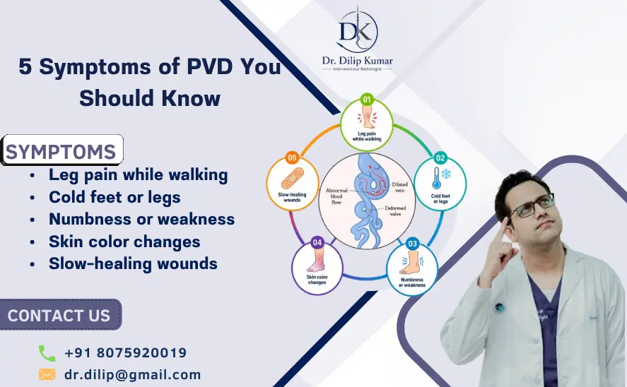
Peripheral Vascular Disease (PVD), often used interchangeably with peripheral artery disease (PAD) when referring to arterial circulation, occurs when blood vessels outside the heart and brain become narrowed or blocked. In the legs, reduced blood flow can cause pain, weakness, skin changes, slow-healing wounds, and other symptoms. Some people may have few or no obvious symptoms, which makes awareness and timely evaluation important.
5 Symptoms of PVD You Should Know can help you recognize changes that should not be dismissed as normal ageing, muscle fatigue, or arthritis. Early assessment may help identify reduced circulation before the condition becomes more serious.
The American Heart Association recommends evaluating people with suspected PAD through medical history, physical examination, and appropriate vascular testing.
5 Symptoms of PVD You Should Know
Peripheral vascular disease can affect the arteries that supply blood to the legs and feet. The most common cause of lower-extremity PAD is atherosclerosis, in which fatty deposits build up inside the arteries and restrict blood flow. Risk is higher with factors such as diabetes, smoking, high blood pressure, high cholesterol, chronic kidney disease, and older age.
One of the most important reasons to understand 5 Symptoms of PVD You Should Know is that symptoms can be subtle at first. A person may notice discomfort only after walking a certain distance and assume it is simply due to age or lack of exercise.
However, recurring leg symptoms—particularly those that appear during activity and improve with rest—deserve medical attention.
What causes PVD?
PVD can develop when arteries become narrowed or blocked. Atherosclerosis is a major cause, but other vascular conditions can also contribute.
Common risk factors include:
- Smoking or tobacco use
- Diabetes
- High blood pressure
- High cholesterol
- Chronic kidney disease
- Increasing age
- A history of cardiovascular disease
- Limited physical activity
- A family history of vascular disease
Because PAD is also associated with an increased risk of heart attack and stroke, diagnosis is not only about treating leg symptoms. It is also an opportunity to identify and manage broader cardiovascular risk.
1. Leg pain or cramping while walking
One of the classic 5 Symptoms of PVD You Should Know is pain, aching, cramping, burning, or fatigue in the leg during walking or exercise.
This is commonly called intermittent claudication. It may occur in the calf, thigh, buttock, hip, or foot, depending on where the arterial narrowing is located. The discomfort typically improves after stopping and resting.
Leg Pain Due to PVD happens because active muscles require more oxygen-rich blood. When an artery is narrowed, blood supply may not increase sufficiently during exercise, resulting in discomfort.
A particularly important pattern is pain that repeatedly begins after walking a similar distance and improves within several minutes of rest.
2. Cold feet or one leg feeling cooler
Another of the 5 Symptoms of PVD You Should Know is an unusual difference in temperature between the two legs or feet.
Reduced arterial circulation may cause one foot or lower leg to feel noticeably cooler than the other. This can be particularly concerning when accompanied by pain, numbness, color changes, or reduced pulses.
Cold feet can have many causes, so this symptom alone does not confirm PVD. However, persistent or one-sided temperature changes should be evaluated, especially in people with diabetes, smoking history, or other vascular risk factors.
3. Slow-healing wounds on the feet or toes
The third of the 5 Symptoms of PVD You Should Know is a wound, blister, cut, or ulcer that takes unusually long to heal.
Adequate blood flow is essential for delivering oxygen and nutrients needed for tissue repair. Significant arterial disease can therefore interfere with wound healing.
A persistent wound on the toe, foot, or lower leg deserves prompt medical evaluation, particularly if there is diabetes or known vascular disease. The AHA notes that nonhealing foot or toe wounds are among the possible signs of PAD.
4. Skin, hair, or toenail changes
The fourth of the 5 Symptoms of PVD You Should Know involves visible changes in the legs and feet.
Possible changes include:
- Pale or unusually red skin
- Reduced hair growth on the legs
- Slow-growing toenails
- Thinner-looking skin
- Loss of calf muscle in advanced disease
- Changes in skin temperature
The skin may appear paler when the leg is elevated and become reddish when lowered. These findings can occur when arterial blood supply is reduced.
5. Numbness, weakness, or pain at rest
The fifth of the 5 Symptoms of PVD You Should Know is numbness, weakness, or pain that occurs even when you are not walking.
Early PAD often produces symptoms during activity. More advanced disease can cause discomfort at rest, particularly in the foot or toes. Severe circulation problems may also be associated with tissue damage, nonhealing wounds, or gangrene.
Rest pain or a sudden major change in circulation should never be ignored. Sudden severe leg pain, marked coldness, pale or blue skin, or sudden loss of sensation or movement can indicate acute limb ischemia and require emergency medical attention.
Diagnosis and Treatment of PVD
Recognizing 5 Symptoms of PVD You Should Know is only the first step. A healthcare professional must determine whether symptoms are actually caused by PAD or another condition, such as nerve compression, arthritis, muscle problems, or other circulation disorders.
How is PVD diagnosed?
Diagnosis generally begins with a detailed medical history and physical examination. The clinician may check pulses in the legs and feet, examine the skin, and ask when symptoms occur.
An important first-line test is the ankle-brachial index (ABI). It compares blood pressure at the ankle with blood pressure in the arm. An ABI of 0.90 or lower is considered abnormal, while values above 1.40 may indicate noncompressible arteries and require additional assessment.
Depending on the findings, additional tests may include:
- Doppler or duplex ultrasound
- Exercise ABI
- CT angiography
- MR angiography
- Catheter angiography
- Toe-brachial index or other perfusion tests in selected patients
The 2024 ACC/AHA guideline recommends using vascular imaging such as duplex ultrasound, CTA, MRA, or catheter angiography when anatomical information is needed to plan revascularization.
Treatment options
Treatment depends on the severity of disease, symptoms, artery involved, overall health, and risk of limb complications.
Management may include:
- Smoking cessation
- Structured exercise
- Healthy diet and weight management
- Cholesterol-lowering treatment
- Blood-pressure control
- Diabetes management
- Antiplatelet or other appropriate antithrombotic medication
- Treatment of other cardiovascular risk factors
Structured exercise is an important part of PAD care. If symptoms remain functionally limiting despite appropriate medical therapy and exercise, revascularization may be considered.
Minimally invasive vascular procedures
For selected patients, an endovascular procedure may help restore blood flow. Depending on the blockage, an Interventional Radiologist in Kannur may use angioplasty, stenting, thrombectomy, or other catheter-based techniques.
The procedure generally involves guiding a thin catheter through a blood vessel to the area of narrowing or blockage. A balloon may be used to widen the narrowed segment, while a stent may be placed when clinically appropriate.
These procedures are not necessary for every patient with PVD. The decision should be based on symptoms, disease severity, imaging findings, treatment response, and individual risks.
Benefits of appropriate PVD treatment
Effective treatment can help:
- Reduce walking-related leg discomfort
- Improve walking ability and physical function
- Improve blood flow
- Support wound healing when poor circulation is contributing
- Improve quality of life
- Reduce the risk of serious limb complications in appropriately selected patients
The 2024 guideline recommends revascularization to prevent limb loss in chronic limb-threatening ischemia and considers it appropriate for selected patients with lifestyle-limiting claudication that has not responded adequately to medical therapy and structured exercise.
Recovery after minimally invasive treatment
Recovery varies according to the procedure and the patient’s health. Catheter-based vascular procedures generally involve less invasive access than traditional open surgery, but the exact recovery period depends on the treatment performed.
Patients may be advised to:
- Keep the access site clean and monitor it for complications
- Follow prescribed medications
- Gradually return to normal activity as advised
- Continue walking or structured exercise when recommended
- Control blood pressure, cholesterol, and diabetes
- Avoid tobacco
- Attend follow-up appointments
Long-term management remains important because treating a blockage does not remove the underlying risk factors for vascular disease.
PVD Treatment in Kannur: When to See an Interventional Radiologist
If you are experiencing 5 symptoms of PVD You Should Know, particularly recurring Leg Pain Due to PVD, unexplained foot wounds, or changes in circulation, consider getting a vascular assessment rather than waiting for symptoms to worsen.
Dr. Dilip Kumar is a Senior Consultant in Endo Vascular & Neuro Interventional Radiology at Aster MIMS Kannur. His listed areas of expertise include peripheral vascular procedures such as mesenteric ischemia stenting, as well as vascular interventions including acute DVT thrombolysis, thrombectomy, stenting, and IVC filter placement.
His interventional radiology practice includes minimally invasive vascular procedures designed to diagnose and treat selected blood-vessel conditions. The appropriate procedure depends on the patient’s imaging findings and clinical condition.
For patients with suspected peripheral arterial disease, consultation can help determine whether symptoms are related to reduced arterial circulation and whether lifestyle management, medication, exercise therapy, or an endovascular intervention may be appropriate.
PVD vs other causes of leg pain
| Feature | PVD-related pain | Muscle/Joint pain | Nerve-related pain |
| Typical trigger | Walking or exercise | Movement or specific activity | Position, movement, or nerve irritation |
| Relief | Often improves with rest | Depends on the cause | May change with position |
| Location | Calf, thigh, buttock, foot | Muscle or joint area | Often follows a nerve pattern |
| Other signs | Coldness, color changes, weak pulses | Swelling/tenderness may occur | Tingling, burning, numbness |
| Evaluation | ABI and vascular imaging | Physical examination/imaging | Neurological assessment/imaging |
Symptoms can overlap, so self-diagnosis is unreliable. A vascular evaluation is particularly important when symptoms repeatedly occur during walking or are accompanied by skin changes or nonhealing wounds.
FAQs About PVD Symptoms
1. What are the most common Peripheral Vascular Disease (PVD) Symptoms?
The most common symptom of lower-extremity PAD is pain, cramping, aching, or fatigue during walking or exercise that improves with rest. Other symptoms include cold feet, skin changes, slow-healing wounds, and reduced hair or nail growth.
2. Is leg pain always caused by PVD?
No. Leg pain can result from arthritis, muscle problems, nerve compression, spinal conditions, injuries, or other disorders. The pattern of pain and appropriate vascular testing help determine the cause.
3. What is Leg Pain Due to PVD Like?
Leg Pain Due to PVD commonly appears during walking or exercise and improves after resting. It may feel like cramping, aching, burning, discomfort, or muscle fatigue.
4. Can PVD occur without symptoms?
Yes. Some people have PAD without obvious symptoms. This is one reason risk assessment and appropriate vascular evaluation are important in people who have significant risk factors.
5. Is PVD dangerous?
PVD can become serious, particularly when blood flow is severely reduced. PAD is also associated with a higher risk of cardiovascular events, including heart attack and stroke.
6. What test is commonly used for PVD?
The ankle-brachial index, or ABI, is a commonly used non-invasive test. Additional ultrasound or vascular imaging may be recommended depending on the findings.
7. Can PVD be treated without surgery?
Yes. Depending on severity, treatment can include exercise, smoking cessation, medication, and management of cardiovascular risk factors. Some patients with significant symptoms may require minimally invasive revascularization.
8. When should I see a doctor for PVD symptoms?
See a healthcare professional if you repeatedly develop leg pain while walking, unexplained foot wounds, changes in skin color or temperature, numbness, or other persistent circulation-related symptoms.
9. When is PVD an emergency?
Sudden severe leg pain with a cold, pale or blue limb, sudden numbness, weakness, or loss of movement may indicate an acute circulation problem. Seek emergency medical care immediately.
10. Can PVD symptoms improve after treatment?
Many patients can experience improvement in symptoms and walking ability with appropriate treatment. The best approach depends on the severity and location of arterial disease and the individual’s overall health.
Conclusion: 5 Symptoms of PVD You Should Know
Understanding 5 Symptoms of PVD You Should Know can help you recognize potential circulation problems before they significantly affect your mobility or quality of life. Recurring walking-related pain, cold feet, slow-healing wounds, skin or nail changes, and pain or numbness at rest should not be automatically attributed to aging.
The good news is that PVD can be evaluated with established vascular tests, and treatment can range from lifestyle and medication management to minimally invasive revascularization for appropriately selected patients.
If you have 5 Symptoms of PVD You Should Know, especially persistent Leg Pain Due to PVD, seeking an early vascular evaluation can help identify the cause and determine the most suitable treatment approach.
Book an appointment with Dr. Dilip Kumar, Interventional Radiologist in Kannur, for expert evaluation of peripheral vascular problems and minimally invasive vascular treatment options. His Aster MIMS profile lists extensive endovascular and interventional radiology expertise, including vascular stenting, thrombectomy and thrombolysis procedures.